
Case report. Swallowed fishbone in an emergency setting and imaging findings
Keywords:
foreign body, soft tissue neck, x-rayAbstract
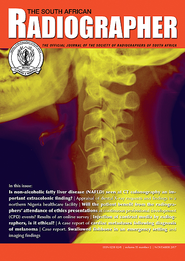
A 27-year-old woman was admitted to the emergency department after swallowing a fish bone while eating dinner. A lateral neck radiograph was performed and a fishbone was detected as a fairly-distinct linear radiopaque density in the pre-cervical soft tissues at the level of the C4/C5 vertebral bodies.
Â
Downloads
Published
2017-12-04
Issue
Section
Case Reports
License
Copyright on all published material belongs to the Society of Radiographers of South Africa (SORSA).I hereby understand and declare that:
- All proprietary rights other than copyright are reserved to the authors, as well as the right to reproduce original figures and tables from this item in their future works, provided full credit is given to the original publication The South African Radiographer ISSN 0258 0241.
- In consideration of the reviewing and editing done by the editors of The South African Radiographer of the above named manuscript, the author/s hereby transfer, assign, or otherwise convey all copyright ownership world-wide, in all languages, to the Society of Radiographers of South Africa in the event that this manuscript is accepted for publication.
- If the manuscript has been commissioned by another person or organisation, or if it has been written as part of the duties of an employee, that full authorization has been given by the representative of the commissioning organisation or employer to be published in the The South African Radiographer.
